BPA (Bisphenols)
At a Glance
Bisphenols are endocrine-disrupting compounds and reproductive toxicants. Bisphenols such as BPA, BPF, BPS, and BPAF are some of the most common chemicals we’re exposed to each day, and they are in everything from food and drink containers to dental fillings.
BPA can disrupt the hormone system, particularly when exposures occur during prenatal development and infancy. Though fewer in number, studies on other bisphenols (BPS, BPF, BPSF) indicate that they mimic the adverse health effects of BPA in cells.

What is BPA?
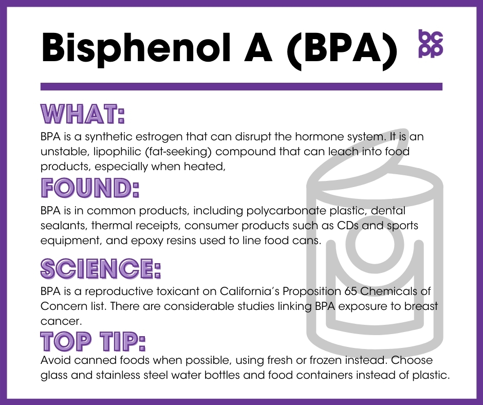
BPA is a synthetic estrogen that can disrupt the hormone system. It is an unstable, lipophilic (fat-seeking) compound that can leach into food products, especially when heated,[1] and food products contaminated with this chemical are thought to be a major source of BPA exposure.[2],[3] Some BPA alternatives, such as bisphenol S (BPS), have come on the market, but have not been proven safe, as they, too, may cause endocrine disruption.[4],[5],[6]
Where is BPA found?
BPA is common in many everyday products, including polycarbonate plastic (widely used in clear plastic containers, eyeglasses, and other household products), dental sealants, thermal receipts, consumer products such as CDs and sports equipment, and epoxy resins used to line food cans.[7],[8] Significant levels have also been measured in ambient air,[9] house dust,[10] and river and drinking water.[11]
BPA can be ingested into the body through food and drink packaged in materials containing BPA. Studies have found rapid increases in BPA levels in urine and blood samples taken from subjects who intentionally increased their intake of food and drinks packaged in BPA-containing products.[12],[13] Another study demonstrated that limiting the intake of commercially packaged food significantly decreased BPA levels in the body.[14]
While food is a major source of BPA, studies of fasting individuals indicate that other sources of BPA exist in everyday environments.[15] BPA has been shown to rapidly absorb into the skin upon brief contact with items such as BPA-containing thermal receipts.[16]
The bisphenol substitutes (BPAF, BPF, BPS) can be found in most of the same types of products described above for BPA, including many products that are labelled as BPA-free.[17]
The US Food & Drug Administration (FDA) banned BPA from baby bottles and infant formula packaging. With the removal of BPA from many products, following both regulatory and market campaign pressures, companies have been replacing BPA with other chemical forms of bisphenol. Unfortunately, research now shows that these substitutes often also act as hormone disruptors, perhaps even more powerfully than BPA.
What evidence links BPA to breast cancer?
BPA is ubiquitous in our environment. It is a reproductive toxicant on California’s Proposition 65 Chemicals of Concern list.[18] Many studies have revealed the accumulating evidence linking BPA exposure to breast cancer
- Although BPA has a rapid clearance rate from the body (hours to days),[19] studies have found it in blood and urine samples of pregnant women,[20],[21],[22] breast milk,[23],[24] amniotic fluid,[25] placental tissue and umbilical cord blood at birth,[27],[26] and the urine of premature infants.[28] This is quite concerning, considering the growing evidence linking BPA to adverse health effects.
- Several studies on rats and mice have found that prenatal exposure to BPA can lead to abnormalities in mammary tissue development. These aberrations were observable during gestation and were maintained into adulthood.[29][30][31][32]
- Many studies using both rat and mouse models have demonstrated that even brief exposures to doses of BPA on par with those measured in everyday settings during gestationor around the time of birth lead to changes in mammary tissue structure predictive of later development of tumors.[33],[34],[35]
- Prenatal exposure of rats to BPA resulted in an increase in pre-cancerous lesions and abnormal cell groups[36],[37] as well as an increased number of mammary tumors following adult exposures to sub-threshold doses of known carcinogens.[38],[39] Later studies demonstrated that BPA alone also increased the growth of mammary tumors, without the addition of known carcinogens.[40]
- Some data suggest that during the time between conception and birth, BPA alters the content and expression of several proteins in some cells, which ultimately modifies gene expression in the mammary gland.[41],[42]
- Studies have shown that the mammary glands of mice exposed to BPA as newborns were more sensitive to estrogen and progesterone during puberty.[43],[44]
- Some of the long-term effects of neonatal exposures to BPA may be dose dependent, with low- and high-dose exposures resulting in different timingand profiles of changes in gene expression in cells of the mammary gland. In one study, low-dose exposures had the most profound effect on rat mammary glands during the period just before reproductive maturity, while higher doses had more delayed effects, altering gene expression in the mammary tissues of mature adults.[45]
- Prenatal exposure of mice to BPA may reduce the immune responses that commonly target developing cancer cells.[46]
- Exposure to low levels of BPA in normal and cancerous human breast cells led to altered gene expression including cell death, rapid increase in cell formation, and the initiation of cancer formation.[47],[48],[49],[50] In the presence of BPA, highly aggressive tumors were developed in cells from the non-cancerous breast of women diagnosed with breast cancer.[51]
- Administration of low doses of BPA led to significantly larger and faster growing mammary tumors in adult mice who had earlier received grafts of undifferentiated cells associated with development of triple negative breast cancer.[52]
- BPA has been found to reduce the effectiveness of common chemotherapy agents (cisplatin, doxirubicin and vinblastin) in blocking the proliferation of human breast cancer cells when tested in vitro.[53],[54] The full implications of these results have not been studied in women.
What evidence links the BPA substitutes BPAF, BPF and BPS to breast cancer?
- In mice, prenatal exposures to BPS or BPAF led to changes in mammary tissue development that were observable at the onset of puberty and which persisted through to adulthood. As young adults, mice treated prenatally with BPS or BPAF showed abnormalities in mammary tissue including increased hyperplasia (abnormal growth of mammary cells), increased inflammation, and higher numbers of tumors. These effects were more profound in mice treated with BPS and BPAF than were found in animals treated with similar doses of BPA.[55]
- Exposure to low levels of BPF in human breast cancer cells led to increases in proliferation and other cellular changes associated with the development of mammary tumors.[56]
- Exposure of human breast cancer cells to low doses of BPAF showed that this substitute activates similar cellular pathways, regulated by the estrogen receptor proteins, as does exposure of the same cells to BPA. Higher doses of BPAF, however, may actually activate pathways that inhibit the development of breast cancers.[57],[58]
- Also in human breast cancer cells, BPS and BPF have been shown to mimic the effects of BPA increasing proliferation of the cells, altering expression of genes involved in regulating the cell cycle in ways associated with development of tumors, and increasing the migration of the cells. These data indicate that BPS and BPF exposures have similar risk profiles as BPA for increasing the development and progression of breast cancer.[59]
Who is most likely to be exposed to BPA?
BPA exposure is widespread and present in many parts of our everyday environment. Individuals who consume foods packaged in cans or use polycarbonate water bottles or baby bottles may have higher exposures. Individuals who work with thermal cash register receipts may absorb BPA through their skin.
Who is most vulnerable to the health of effects of BPA?
Studies on rats and mice have found that BPA exposure during pregnancy has been shown to significantly impact fetal development.[60],[61],[62],[63],[64],[65],[66],[67],[68]
What are the top tips to avoid exposure?
Luckily, clearance of BPA from the body is quite rapid. One study demonstrated that just a three-day period of limiting intake of packaged foods decreased the concentrations of BPA found in urine by an average 65 percent.[69]
Making these simple changes can reduce your exposure:
- Avoid canned foods. Where fresh alternatives are not available, choose frozen.
- Avoid clear, shatterproof plastic food and drink containers (sometimes labeled with the recycling code 7).
- Avoid handling thermal receipts.
- Choose BPA-free baby bottles and child cups. It is important to be aware that the alternatives to BPA have not been adequately tested. Glass and stainless steel containers are your safest bet. BPA-free labels unfortunately do not always mean the product is safe.
Updated 2019
[2] Lakind, Judy S, and Daniel Q Naiman. “Daily intake of bisphenol A and potential sources of exposure: 2005-2006 National Health and Nutrition Examination Survey.” Journal of Exposure Science & Environmental Epidemiology 21, 3 (2011): 272-9. doi:10.1038/jes.2010.9.
[3] Vandenberg, Laura N et al. “Human exposure to bisphenol A (BPA).” Reproductive Toxicology 24, 2 (2007): 139-77. doi:10.1016/j.reprotox.2007.07.010.
[4] Rochester, Johanna R, and Ashley L Bolden. “Bisphenol S and F: A Systematic Review and Comparison of the Hormonal Activity of Bisphenol A Substitutes.” Environmental Health Perspectives 123, 7 (2015): 643-50. doi:10.1289/ehp.1408989.
[5] Ji, Kyunghee, Hong, Seongjin Hong, Younglim Kho and Kyungho Choi. “Effects of bisphenol S exposure on endocrine functions and reproduction of zebrafish.” Environmental Science & Technology 47,15 (2013): 8793-800. doi:10.1021/es400329t.
[6] Kim, Ji-Youn et al. “Effects of bisphenol compounds on the growth and epithelial mesenchymal transition of MCF-7 CV human breast cancer cells.” Journal of Biomedical Research 31,4 (2017): 358-369. doi:10.7555/JBR.31.20160162.
[7] California Office of Environmental Health Hazard Assessment. “Chemicals: Bisphenol-A.” Last modified 2020. http://oehha.ca.gov/chemicals/bisphenol-a.
[8] Xue, Jingchuan et al. “Bisphenols, Benzophenones, and Bisphenol A Diglycidyl Ethers in Textiles and Infant Clothing.” Environmental Science & Technology 51,9 (2017): 5279-5286. doi:10.1021/acs.est.7b00701.
[9] Matsumoto, H, S. Adachi and Y. Suzuki. “Bisphenol A in ambient air particulates responsible for the proliferation of MCF-7 human breast cancer cells and Its concentration changes over 6 months.” Archives of Environmental Contamination and Toxicology 48,4 (2005): 459-66. doi:10.1007/s00244-003-0243-x.
[10] Rudel Ruthann, David E. Camann, John D. Spengler, Leo R. Korn and Julia G Brody. “Phthalates, alkylphenols, pesticides, polybrominated diphenyl ethers, and other endocrine-disrupting compounds in indoor air and dust.” Environmental Science & Technology 37,20 (2003): 4543-53. doi:10.1021/es0264596.
[11] Rodriguez-Mozaz S, ML de Alda and D. Barceló. “Analysis of bisphenol A in natural waters by means of an optical immunosensor.” Water Research 39 (2005): 5071-9. doi:10.1016/j.watres.2005.09.023.
[12] Carwile, Jenny L et al. “Polycarbonate bottle use and urinary bisphenol A concentrations.” Environmental Health Perspectives 117, 9 (2009): 1368-72. doi:10.1289/ehp.0900604.
[13] Smith R, Lourie. Slow Death by Rubber Duck: The Secret Danger of Everyday Things. Berkeley, CA: Counterpoint, 2009.
[14] Rudel, Ruthann A et al. “Food packaging and bisphenol A and bis(2-ethyhexyl) phthalate exposure: findings from a dietary intervention.” Environmental Health Perspectives 119,7 (2011): 914-20. doi:10.1289/ehp.1003170.
[15] Stahlhut, Richard W, Wade V Welshons and Shanna H Swan. “Bisphenol A data in NHANES suggest longer than expected half-life, substantial nonfood exposure, or both.” Environmental Health Perspectives 117,5 (2009): 784-9. doi:10.1289/ehp.0800376.
[16] vom Saal, Frederick S, and Wade V Welshons. “Evidence that bisphenol A (BPA) can be accurately measured without contamination in human serum and urine, and that BPA causes numerous hazards from multiple routes of exposure.” Molecular and Cellular Endocrinology 398,1-2 (2014): 101-13. doi:10.1016/j.mce.2014.09.028.
[17] Rochester, Johanna R, and Ashley L Bolden. “Bisphenol S and F: A Systematic Review and Comparison of the Hormonal Activity of Bisphenol A Substitutes.” Environmental Health Perspectives 123,7 (2015): 643-50. doi:10.1289/ehp.1408989.
[18] California Office of Environmental Health Hazard Assessment. “Chemicals Considered or Listed Under Proposition 65: Bisphenol A (BPA).” Last modified 2020. http://oehha.ca.gov/proposition-65/chemicals/bisphenol-bpa.
[19] Stahlhut, Richard W, Wade V Welshons and Shanna H Swan. “Bisphenol A data in NHANES suggest longer than expected half-life, substantial nonfood exposure, or both.” Environmental Health Perspectives 117,5 (2009): 784-9. doi:10.1289/ehp.0800376.
[20] Ye, Xibiao et al. “Levels of metabolites of organophosphate pesticides, phthalates, and bisphenol A in pooled urine specimens from pregnant women participating in the Norwegian Mother and Child Cohort Study (MoBa).” International Journal of Hygiene and Environmental Health 212, 5 (2009): 481-91. doi:10.1016/j.ijheh.2009.03.004.
[21] Padmanabhan, V et al. “Maternal bisphenol-A levels at delivery: a looming problem?.” Journal of Perinatology: Official Journal of the California Perinatal Association 28,4 (2008): 258-63. doi:10.1038/sj.jp.7211913.
[22] Teeguarden Justin G, Nathan C. Twaddle, Mona I. Churchwell and Daniel R. Doerge. “Urine and serum biomonitoring of exposure to environmental estrogens I: Bisphenol A in pregnant women.” Food and Chemical Toxicology 92 (2016): 129-142. https://doi.org/10.1016/j.fct.2016.03.023.
[23] Kuruto-Niwa Ryoko et al. “Estrogenic activity of the chlorinated derivatives of estrogens and flavonoids using a GFP expression system.” Environmental Toxicology and Pharmacology 23, 1 (2007): 121-128. https://doi.org/10.1016/j.etap.2006.07.011.
[24] Sun, Yen et al. “Determination of bisphenol A in human breast milk by HPLC with column-switching and fluorescence detection.” Biomedical Chromatography: BMC 18,8 (2004): 501-7. doi:10.1002/bmc.345.
[25] Ikezuki, Yumiko et al. “Determination of bisphenol A concentrations in human biological fluids reveals significant early prenatal exposure.” Human Reproduction 17,11 (2002): 2839-41. doi:10.1093/humrep/17.11.2839.
[26] Environmental Working Group. “BPA and Alternatives.” Accessed November 4, 2020. https://www.ewg.org/key-issues/toxics/bpa.
[27] Schönfelder, Gilbert et al. “Parent bisphenol A accumulation in the human maternal-fetal-placental unit.” Environmental Health Perspectives 110,11 (2002): A703-7. doi:10.1289/ehp.110-1241091.
[28] Calafat, Antonia M et al. “Exposure to bisphenol A and other phenols in neonatal intensive care unit premature infants.” Environmental Health Perspectives 117,4 (2009): 639-44. doi:10.1289/ehp.0800265.
[29] Vandenberg, Laura N et al. “Exposure to environmentally relevant doses of the xenoestrogen bisphenol-A alters development of the fetal mouse mammary gland.” Endocrinology 148,1 (2007): 116-27. doi:10.1210/en.2006-0561.
[30] Vandenberg, Laura N et al. “Perinatal exposure to the xenoestrogen bisphenol-A induces mammary intraductal hyperplasias in adult CD-1 mice.” Reproductive Toxicology 26,3-4 (2008): 210-9. doi:10.1016/j.reprotox.2008.09.015.
[31] Betancourt, Angela M et al. “Proteomic analysis in mammary glands of rat offspring exposed in utero to bisphenol A.” Journal of Proteomics 73,6 (2010): 1241-53. doi:10.1016/j.jprot.2010.02.020.
[32] Perrot-Applanat, Martine et al. “Alteration of mammary gland development by bisphenol a and evidence of a mode of action mediated through endocrine disruption.” Molecular and Cellular Endocrinology 475 (2018): 29-53. doi:10.1016/j.mce.2018.06.015.
[33] Markey, C M et al. “In utero exposure to bisphenol A alters the development and tissue organization of the mouse mammary gland.” Biology of Reproduction 65,4 (2001): 1215-23. doi:10.1093/biolreprod/65.4.1215.
[34] Maffini, Maricel V et al. “Endocrine disruptors and reproductive health: the case of bisphenol-A.” Molecular and Cellular Endocrinology 254-255 (2006): 179-86. doi:10.1016/j.mce.2006.04.033.
[35] Muñoz-de-Toro, Monica et al. “Perinatal exposure to bisphenol-A alters peripubertal mammary gland development in mice.” Endocrinology 146,9 (2005): 4138-47. doi:10.1210/en.2005-0340.
[36] Murray, Tessa J et al. “Induction of mammary gland ductal hyperplasias and carcinoma in situ following fetal bisphenol A exposure.” Reproductive Toxicology 23,3 (2007): 383-90. doi:10.1016/j.reprotox.2006.10.002.
[37] Acevedo, Nicole et al. “Perinatally administered bisphenol a as a potential mammary gland carcinogen in rats.” Environmental Health Perspectives 121,9 (2013): 1040-6. doi:10.1289/ehp.1306734.
[38] Durando, Milena et al. “Prenatal bisphenol A exposure induces preneoplastic lesions in the mammary gland in Wistar rats.” Environmental Health Perspectives 115,1 (2007): 80-6. doi:10.1289/ehp.9282.
[39] Jenkins Sarah et al. “Oral exposure to bisphenol a increases dimethylbenzanthracene-induced mammary cancer in rats.” Environmental Health Perspectives 117, 6 (2009): 910-15. doi:10.1289/ehp.11751.
[40] Acevedo, Nicole et al. “Perinatally administered bisphenol a as a potential mammary gland carcinogen in rats.” Environmental Health Perspectives 121,9 (2013): 1040-6. doi:10.1289/ehp.1306734.
[41] Wadia, Perinaaz R et al. “Low-dose BPA exposure alters the mesenchymal and epithelial transcriptomes of the mouse fetal mammary gland.” PloS One 8, 5 (2013): e63902. doi:10.1371/journal.pone.0063902.
[42] Ibrahim, Marwa A A et al. “Effect of bisphenol A on morphology, apoptosis and proliferation in the resting mammary gland of the adult albino rat.” International Journal of Experimental Pathology 97, 1 (2016): 27-36. doi:10.1111/iep.12164.
[43] Wadia, Perinaaz R et al. “Perinatal bisphenol A exposure increases estrogen sensitivity of the mammary gland in diverse mouse strains.” Environmental Health Perspectives 115, 4 (2007): 592-8. doi:10.1289/ehp.9640.
[44] Ayyanan, Ayyakkannu et al. “Perinatal exposure to bisphenol a increases adult mammary gland progesterone response and cell number.” Molecular Endocrinology 25, 11 (2011): 1915-23. doi:10.1210/me.2011-1129.
[45] Moral, Raquel et al. “Effect of prenatal exposure to the endocrine disruptor bisphenol A on mammary gland morphology and gene expression signature.” The Journal of Endocrinology 196,1 (2008): 101-12. doi:10.1677/JOE-07-0056.
[46] Fischer, Catha et al. “Bisphenol A (BPA) Exposure In Utero Leads to Immunoregulatory Cytokine Dysregulation in the Mouse Mammary Gland: A Potential Mechanism Programming Breast Cancer Risk.” Hormones & Cancer 7,4 (2016): 241-51. doi:10.1007/s12672-016-0254-5.
[47] Goodson, William H 3rd et al. “Activation of the mTOR pathway by low levels of xenoestrogens in breast epithelial cells from high-risk women.” Carcinogenesis 32, 11 (2011): 1724-33. doi:10.1093/carcin/bgr196.
[48] Tilghman, Syreeta L et al. “Endocrine disruptor regulation of microRNA expression in breast carcinoma cells.” PloS One 7,3 (2012): e32754. doi:10.1371/journal.pone.0032754.
[49] Weng, Yu-I et al. “Epigenetic influences of low-dose bisphenol A in primary human breast epithelial cells.” Toxicology and Applied Pharmacology 248, 2 (2010): 111-21. doi:10.1016/j.taap.2010.07.014.
[50] Xu F et al. “Bisphenol A induces proliferative effects on both breast cancer cells and vascular endothelial cells through a shared GPER-dependent pathway in hypoxia.” Environmental Pollution 231 (2017):1609-1620. doi: 10.1016/j.envpol.2017.09.069.
[51] Dairkee, Shanaz H et al. “Bisphenol A induces a profile of tumor aggressiveness in high-risk cells from breast cancer patients.” Cancer Research 68, 7 (2008): 2076-80. doi:10.1158/0008-5472.CAN-07-6526.
[52] Xu F et al. “Bisphenol A induces proliferative effects on both breast cancer cells and vascular endothelial cells through a shared GPER-dependent pathway in hypoxia.” Environmental Pollution 231 (2017):1609-1620. doi: 10.1016/j.envpol.2017.09.069.
[53] Lapensee, Elizabeth W et al. “Bisphenol A at low nanomolar doses confers chemoresistance in estrogen receptor-alpha-positive and -negative breast cancer cells.” Environmental Health Perspectives 117, 2 (2009): 175-80. doi:10.1289/ehp.11788.
[54] LaPensee, Elizabeth W et al. “Bisphenol A and estradiol are equipotent in antagonizing cisplatin-induced cytotoxicity in breast cancer cells.” Cancer Letters 290, 2 (2010): 167-73. doi:10.1016/j.canlet.2009.09.005.
[55] Tucker, Deirdre K et al. “Evaluation of Prenatal Exposure to Bisphenol Analogues on Development and Long-Term Health of the Mammary Gland in Female Mice.” Environmental Health Perspectives 126, 8 (2018): 087003. doi:10.1289/EHP3189.
[56] Lei, Bingli et al. “Low-concentration BPF induced cell biological responses by the ERα and GPER1-mediated signaling pathways in MCF-7 breast cancer cells.” Ecotoxicology and Environmental Safety 165 (2018): 144-152. doi:10.1016/j.ecoenv.2018.08.102.
[57] Okazaki, Hiroyuki et al. “Bisphenol AF as an Inducer of Estrogen Receptor β (ERβ): Evidence for Anti-estrogenic Effects at Higher Concentrations in Human Breast Cancer Cells.” Biological & Pharmaceutical Bulletin 40, 11 (2017): 1909-1916. doi:10.1248/bpb.b17-00427.
[58] Okazaki, Hiroyuki et al. “Bisphenol AF as an activator of human estrogen receptor β1 (ERβ1) in breast cancer cell lines.” The Journal of Toxicological Sciences 43,5 (2018): 321-327. doi:10.2131/jts.43.321.
[59] Kim, Ji-Youn et al. “Effects of bisphenol compounds on the growth and epithelial mesenchymal transition of MCF-7 CV human breast cancer cells.” Journal of Biomedical Research 31,4 (2017): 358-369. doi:10.7555/JBR.31.20160162.
[60] Maffini, Maricel V et al. “Endocrine disruptors and reproductive health: the case of bisphenol-A.” Molecular and Cellular Endocrinology 254-255 (2006): 179-86. doi:10.1016/j.mce.2006.04.033.
[61] Muñoz-de-Toro, Monica et al. “Perinatal exposure to bisphenol-A alters peripubertal mammary gland development in mice.” Endocrinology 146,9 (2005): 4138-47. doi:10.1210/en.2005-0340.
[62] Vandenberg, Laura N et al. “Perinatal exposure to the xenoestrogen bisphenol-A induces mammary intraductal hyperplasias in adult CD-1 mice.” Reproductive Toxicology 26,3-4 (2008): 210-9. doi:10.1016/j.reprotox.2008.09.015.
[63] Betancourt, Angela M et al. “Proteomic analysis in mammary glands of rat offspring exposed in utero to bisphenol A.” Journal of Proteomics 73,6 (2010): 1241-53. doi:10.1016/j.jprot.2010.02.020.
[64] Murray, Tessa J et al. “Induction of mammary gland ductal hyperplasias and carcinoma in situ following fetal bisphenol A exposure.” Reproductive Toxicology 23,3 (2007): 383-90. doi:10.1016/j.reprotox.2006.10.002.
[65] Acevedo, Nicole et al. “Perinatally administered bisphenol a as a potential mammary gland carcinogen in rats.” Environmental Health Perspectives 121,9 (2013): 1040-6. doi:10.1289/ehp.1306734.
[66] Wadia, Perinaaz R et al. “Perinatal bisphenol A exposure increases estrogen sensitivity of the mammary gland in diverse mouse strains.” Environmental Health Perspectives 115, 4 (2007): 592-8. doi:10.1289/ehp.9640.
[67] Ayyanan, Ayyakkannu et al. “Perinatal exposure to bisphenol a increases adult mammary gland progesterone response and cell number.” Molecular Endocrinology 25, 11 (2011): 1915-23. doi:10.1210/me.2011-1129.
[68] Tucker, Deirdre K et al. “Evaluation of Prenatal Exposure to Bisphenol Analogues on Development and Long-Term Health of the Mammary Gland in Female Mice.” Environmental Health Perspectives 126, 8 (2018): 087003. doi:10.1289/EHP3189.
[69] Rudel, Ruthann A et al. “Food packaging and bisphenol A and bis(2-ethyhexyl) phthalate exposure: findings from a dietary intervention.” Environmental Health Perspectives 119,7 (2011): 914-20. doi:10.1289/ehp.1003170.


