Ionizing Radiation
Ionizing Radiation
Ionizing radiation is an established risk factor for breast cancer. Minimizing radiation dose to breast tissue is critically important, particularly in girls and young women.Science Summary
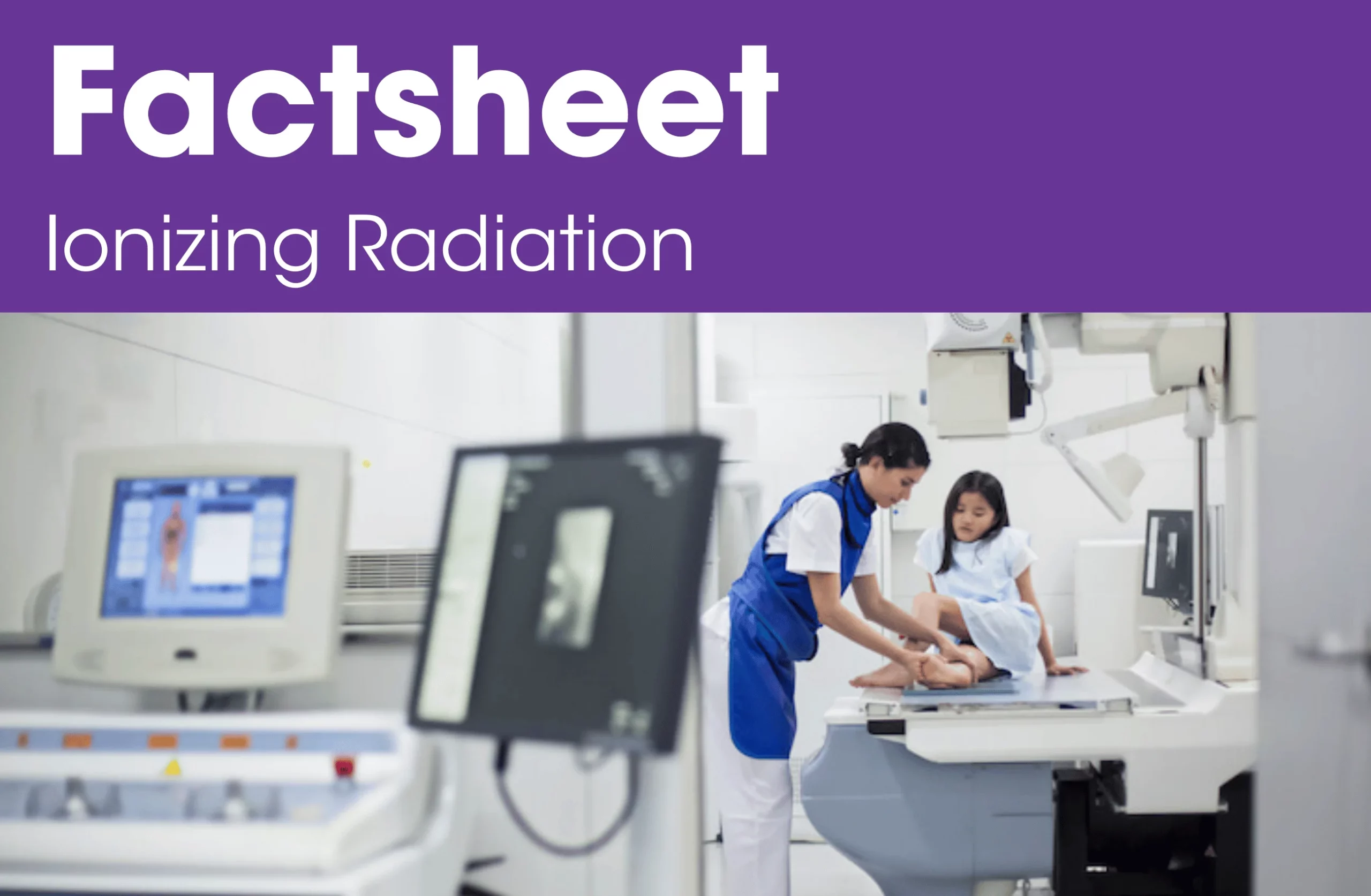
For most people, the largest source of exposure to ionizing radiation is from medical diagnostic procedures, which include x-rays, mammograms, CT scans, and fluoroscopy. Currently, only mammography undergoes close federal oversight. CT scans are of the greatest concern because of their higher doses and frequent use. In the U.S. one CT scan is performed per four individuals and the radiation levels at these examinations are up to 500 times the level of an x-ray radiograph.

For some people, other sources of ionizing radiation exposure include air travel. Work in the nuclear industry, as well as exposure to nuclear weapons production, use, and testing, affects military personnel, civilians, and nearby communities.
Radiation not only increases cancer risk but can also interact with, trigger, or amplify, the impacts of other breast cancer risk factors. These include tobacco, chemicals or chemotherapeutic agents, and individual factors such as age at exposure, gender or reproductive history.
What can I do for my own body and health?
While some exposure to radiation is unavoidable, unnecessary exposures can be eliminated.
Many exposures are medical necessities, however other procedures may be unnecessary. When it comes to medical imaging, ask your doctors and radiation experts to help reduce your exposure to and risk of harm from diagnostic ionizing radiation. Ask that they:

Check if you have a similar test done recently that can provide them with the background information they need.

Check if a test that does not use ionizing radiation (for example, ultrasound or MRI) can provide similar information.

Make certain the least possible amount of radiation needed to obtain a good-quality image is used for your procedure.
What can I do to support the health of my family and friends, and my community?
Share what you know with loved ones. For those who are uncomfortable or nervous making requests to medical professionals, offer to go with them to appointments or practice asking questions at home.
How can I navigate and get support with any systemic barriers to my health?
The University of California-San Francisco offers resources to help understand and reduce medical radiation exposure. Visit knowyourdose.ucsf.edu for more information and tips on discussing dose minimization with your healthcare providers.
People who work in healthcare facilities also face higher exposure to ionizing radiation. It is important for workers to know the risks of medical imaging, and receive training on appropriate use and protection.
In terms of environmental exposures, radon is a naturally occurring radioactive gas and levels in your home can vary depending on where you live and how your house was built. The Environmental Protection Agency’s Radon Zone Map provides information on radon levels. The California Department of Public Health has tips on how to get houses tested and the Environmental Protection Agency has resources on what to do if radon is detected.
How can I help advocate for and support systemic change to remove barriers to health?
Ionizing radiation definitively increases breast cancer risk and it is critical that we reduce this exposure whenever possible. Here are some systems-level changes we can advocate for:

Ensure all medical radiological equipment complies with state standards to minimize radiation exposures from medical imaging.

Establish best practices and train all personnel potentially exposed in health care settings (including, for example, cleaning and other support staff) on how to minimize radiation exposure from medical devices.

Implement best practices to minimize cumulative exposure to radiation in non-medical workplaces.

Require full transparency to residents, developers, and other interested parties of potential radioactive contamination in their communities.
While standards should ultimately be set at the federal level, California has acted to limit exposure to medical radiation. In 2005, the state enacted Assembly Bill 929 (AB 929), Quality Assurance for Radiological Equipment, to require the California Department of Health Services to adopt quality assurance standards that include testing on all radiation-emitting equipment to ensure that the lowest possible dose of radiation is used without sacrificing imaging quality.
Resources
Chapter Pullout

Supported by grant funding from Gilead Sciences, Inc. Gilead Sciences, Inc. has had no input into the development or content of these materials.
Types: Fact Sheet, Report
Related Resources

Genetics
Cancer cells behave differently than normal, healthy cells and tissues. For a long time, cancer was thought to occur because...In California, Cleaning Product Companies to Come Clean on Ingredients
Until now, consumers, workers, and public health agencies have been left in the dark about the potential presence of allergens,...
Tobacco
Tobacco products are full of chemicals that harm our health. We can reduce our risk of breast cancer by avoiding...